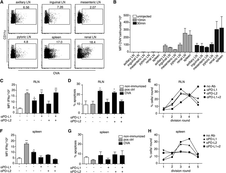
Figure 1.
PD-L1 mediates CTL apoptosis in the renal LN, but not in the spleen. (A) Uptake of OVA-Alexa647 by CD11c+ DCs in various LNs and the spleen 10 minutes after intravenous injection assessed by flow cytometry. Numbers represent the percentage of cells that had captured OVA-Alexa647. (B) Quantitative analysis of OVA uptake in uninjected mice (white) 10 minutes (gray) and 30 minutes (black) after OVA-Alexa647 injection. The absolute numbers of OVA+ DCs was multiplied with their mean fluorescence intensity (MFI) at 647 nm, which correlates with the amount of OVA captured per OVA+ DC. (C–H) MFI of IFN-γ production (C and F) and apoptosis of OT-I cells (D and G) in the RLN (C and D) and the spleen (F and G) 3 days after injecting 8 µg/g body weight endotoxin-free OVA (black), compared with nonimmunized control (white) and immunogenic immunization with OVA/CpG subcutaneously followed by analysis of the draining skin LN (gray). Antibodies blocking PD-L1 and/or PD-L2 are administered intraperitoneally 1 day before and on the day of immunization. (E and H) Proliferation of OT-I cells 3 days after OVA injection in the RLN (E) and the spleen (H) are expressed as proportion of cells per division round, after treatment with blocking antibodies against PD-L1 (triangle), PD-L2 (reverse triangle), or both (square) or left untreated (circle). Data are given as means of three independent experiments. P values refer to nonimmunized controls.

