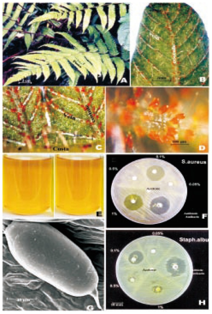
Figure 1. Detailed micromorphological, phytochemical and bioactivity studies on C. parasitica.

A: Leaves of C. parasitica (L.) H. Lev.; B: A pinna lobc showing orange coloured glands on the veins; C: Portion of a pinna lobe with orange coloured glands on cosutules and veins; D: Portion of a vein with cluster of orange coloured glands; E: Glandular extract in acetone; G: Scanning elcctron microscopic view of a gland; F&H: Antibacterial activity of glandular extract against S. aureus (F) and S. albus (H).
