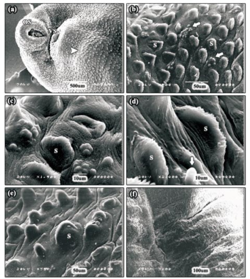
Figure 2. SEMs of adult F. gigantica following 24 h incubation in 30 µg/mL Mirazid®.
a: SEM of the apical cone region showing moderate swelling of the tegumental surface. In the majority of the specimens examined, an often large area of tegumental swelling was observed just posterior to the ventral sucker in the anterior mid-body (head arrow). OS: oral sucker, VS: ventral sucker. b, c: SEMs of the swollen tegument along the apical cone region revealing clusters of normal sensory papillae (arrows). S: spine. d: High power SEM of the tegumental surface, in some specimens, showing severe tegumental swelling. The spines and some sensory papillae (arrow) appeared to be swollen. S: spine. e: SEM of the posterior mid-body region showing moderately swollen tegument and the spines were partially submerged. S: spine. f: SEM of the tail region. In this specimen, the tegument was severely swollen to the extent that the spines were not visible.

