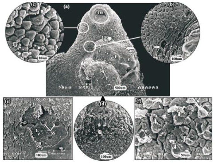
Figure 6. SEMs of adult F. gigantica following 24 h incubation in 60 µg/mL myrrh volatile oil.
a: SEM of the apical cone region showing more pronounced tegumental disruption, in the form of huge swelling (arrow) accompanied with erosions (E), on the ventral surface between the gonopore and the ventral sucker. OS: oral sucker, VS: ventral sucker, GP: gonopore. b: SEM of the tegument close to the gonopore showing small holes (arrow) which penetrated the basal lamina in areas where tegument had been removed. c: SEM of the lateral margin of the apical cone. In this specimen, the tegument was severely swollen and large areas were fissured and accompanied by blebbing (head arrows). S: spine. d: SEM of the anterior mid-body region, directly behind the ventral sucker. in this area, the tegument swelling had become so severe that the spines were barely visible. e: SEM of the posterior mid-body region showing patches of tegumental sloughing (L) and empty spine sockets (SS). f: SEM of the tail region showing mild furrowing of the tegumental syncytium. The tegument covering the spines was seen to be partially sloughed off (arrows).

