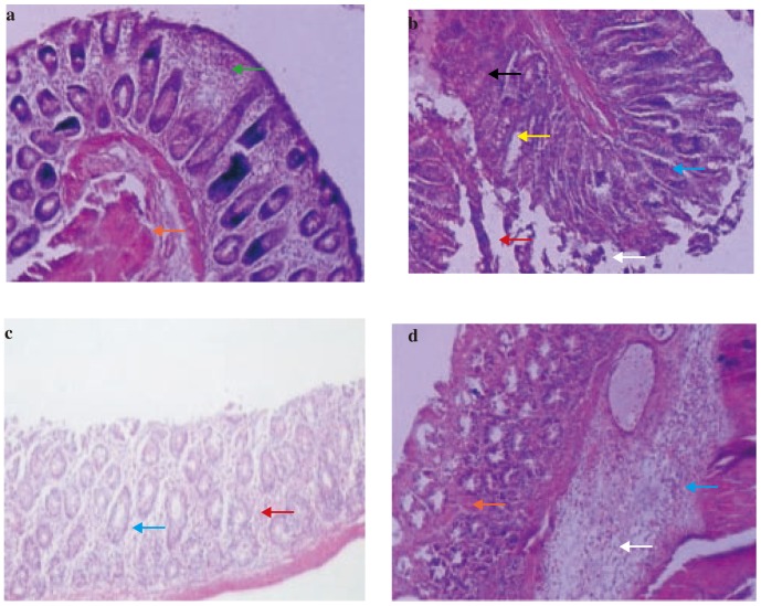
Figure 3. Photomicrographs of sections of colons from rats stained with H&E.
Colon microscopic image of (a) Normal rat with intact epithelial (orange arrow) and mucosal layer (green arrow); (b) Acetic acid induced colitis rat with extensive damage including edema in submucosa (white arrow) and cellular infiltration (blue arrow), hemorrhages (red arrow), necrosis (yellow arrow) and ulceration (black arrow); (c) Prednisolone (2 mg/kg, p.o.) treated rat with infiltration (blue arrow) and hemorrhages (red arrow); (d) HRS (200 mg/kg p.o.) 7 days pretreated rat with edema in submucosa (white arrow), cellular infiltration (blue arrow) and hemorrhages (red arrow). Images (× 100 magnification) are typical and representative of each study group.

