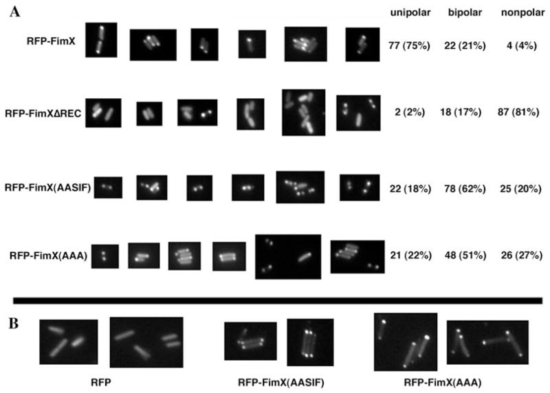
Fig. 6.
Localization of RFP–FimX fusion proteins by indirect immunofluorescence. ΔfimX (panel A) or PA103 (panel B) bacterial colonies expressing indicated RFP–FimX fusion constructs were picked from 2-day-old LB agar plates, resuspended in a drop of ddH2O and spotted onto slides coated with a thin cushion of 1% Gel-Gro gellan. Bacteria were visualized and photographed as detailed in Experimental procedures.

