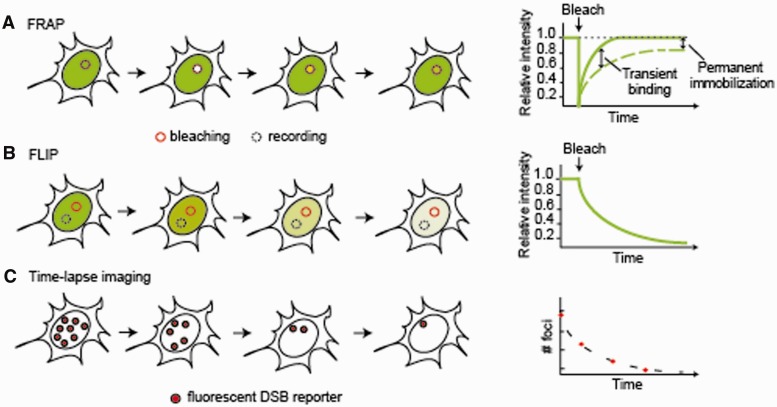
Figure 2:
Live-cell imaging techniques for studying DNA repair. (A) FRAP. A region of interest (solid circle) in the nucleus is photobleached with a strong laser pulse and the recovery of fluorescence intensity within the same region is monitored in time. The diffusion of the tagged protein and the kinetics of its binding interactions are measured from the rate of recovery (dashed line) in comparison to an inert protein of similar size (solid line). A delayed FRAP recovery indicates transient binding events and the fraction of recovery reflects the proportion of mobile molecules. In this example, ∼80% of tagged proteins in the bleached region are mobile. (B) FLIP. Bleaching within a region of interest (solid circle) is accompanied by measurement of fluorescence intensity in another region of the nucleus (dashed circle). The fluorescence decay in the recorded region indicates the extent of exchange between the two compartments. (C) Time-lapse microscopy. The expression and localization of the tagged proteins is monitored in time. In this example, the number of repair foci formed by the tagged protein is monitored to measure the rates of repair.

