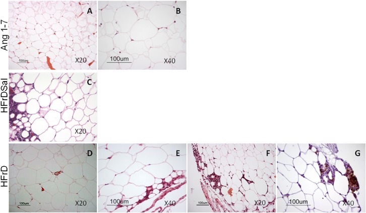
FIG. 3.
Histological sections of epididymal fat tissue. Tissue sections from Ang 1-7–treated rats (n = 6) (A and B) display many small adipocytes and fewer large adipocytes (a field “enriched” with large adipocytes is shown in B). The large adipocytes did not display inflammatory infiltration. Tissue from HFrD and HFrDSal rats (n = 9) (C–G) displays large adipocytes with inflammatory infiltration (C, E, and F), mostly of macrophages (CD68 staining) (G). um, μm.

