
Figure 5.
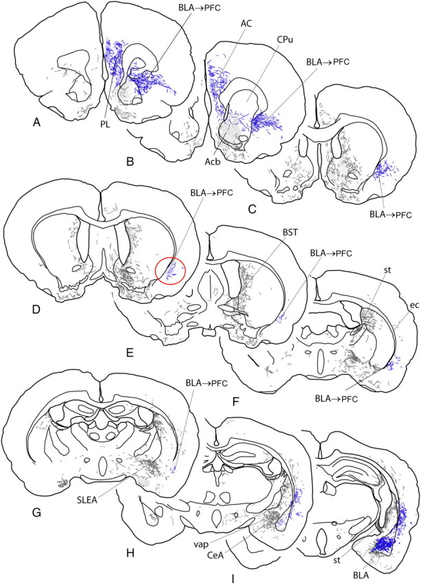
A–I, Diagrams of sections of rat brain ordered from rostral to caudal levels and indicating anterograde labeling produced by an injection of the anterograde tracer biotinylated dextran amine into the BLA, represented by the blue dots in I. Labeled fibers are represented by short lines, which indicate directional orientation, and areas with dense terminal-like labeling are indicated by fine stippling. The pathway from the BLA to the medial prefrontal cortex is highlighted in blue and the site where bupivacaine was infused in the present study to inactivate the pathway is circled in red (D). AC, Anterior cingulate cortex; Acb, nucleus accumbens; BST, bed nucleus of stria terminalis; CeA, central nucleus of the amygdala; CPu, caudate–putamen; ec, external capsule; PL, prelimbic cortex; SLEA, sublenticular extended amygdala; st, stria terminalis; vap, ventral amygdalofugal pathway.