
Figure 3.
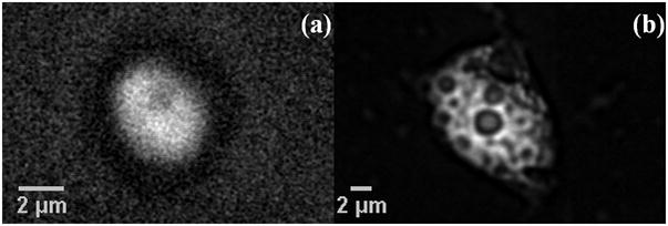
(a) 3-dimensional movie obtained by focusing from top (frame shown) to bottom in different planes in steps of 0.25 μm inside a C. parvum oocyst. (b) F-CARS image of a non-viable cyst of the protozoan pathogen, Giardia Lamblia at 2845 cm−1.
Official websites use .gov
A
.gov website belongs to an official
government organization in the United States.
Secure .gov websites use HTTPS
A lock (
) or https:// means you've safely
connected to the .gov website. Share sensitive
information only on official, secure websites.
(a) 3-dimensional movie obtained by focusing from top (frame shown) to bottom in different planes in steps of 0.25 μm inside a C. parvum oocyst. (b) F-CARS image of a non-viable cyst of the protozoan pathogen, Giardia Lamblia at 2845 cm−1.