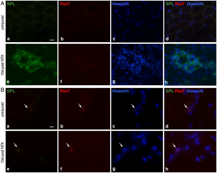
Fig. 5.
SPL is expressed in inflammatory cells of degenerating/regenerating skeletal muscle. Cross-sections of cryopreserved skeletal muscle of an uninjured wild type (C57BL/6) mouse at rest (upper panels) and 10 days post-NTX-injury (bottom panels). Muscles were excised, sectioned, stained with antibodies specific for SPL and Pax7 and counterstained with Hoechst 33342. A: (a to d) resting myofibers do not express SPL; (e to h) strong stain is observed in degenerated/necrotic myofibers undergoing regeneration. Scale bar 100 μm. B: SCs (arrows) that were detected in both injured and uninjured muscles did not co-stain for SPL. Scale bar 10 μm.

