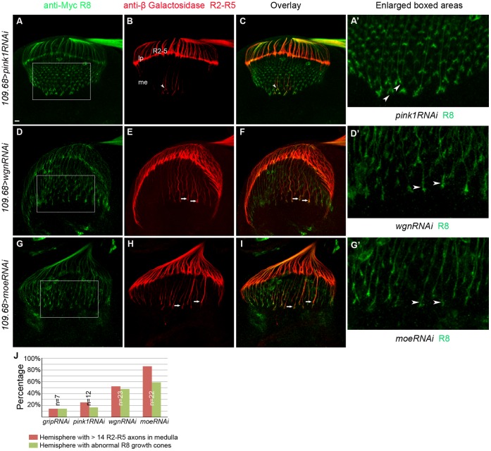
Figure 10. R8-psecific knockdown of Wgn or Moe results in cell-autonomous R8 growth cone defects and non-cell autonomous R2–R5 targeting defects.
Samples were double labeled with Ato-tau-Myc for R8 (green) and Ro-tau-LacZ for R2–R5 (red). (A–C) R8 growth cone morphology and R2–R5 axon targeting were normal in control animals in which unrelated proteins (Pink1 or Grip) were specifically knocked down in R8 cells (w1118/y; 109.68-Gal4/UAS-pink1-RNAi; rtl, atm/+. w1118/y; 109.68-Gal4/ UAS-grip-RNAi; rtl, atm/+). The images showed R8 knock down of Pink1. (D–F) R8-specific knockdown of Wgn resulted in abnormal R8 growth cones (arrow heads in D′) in 47% of hemispheres and caused R2–R5 targeting defects in 56% of hemispheres (arrows in E; w1118/y; 109.68-GALl4/+; UAS-wgn-RNAi/rtl, atm). (G–I) R8-specific knock down of Moe caused R8 growth cone morphology defects (arrow heads in G′) in 64% of hemispheres and R2–R5 mistargeting and/or bundled axons (arrows in H) in 86% of hemispheres (w1118/y; 109.68-Gal4/UAS-moe-RNAi; rtl, atm/+). (A′, D′ and G′) Magnified views in boxed areas in A, D and G. (J) Quantifications. Hemispheres with > 14 R2–R5 axons and/or with thicker bundle in the medullar region were counted as hemispheres with R2–R5 mistargeting defects. Hemispheres that have irregular array of R8 growth cones in the medullar region with blunt or enlarged ends were counted as hemispheres with abnormal R8 growth cones. Abbreviations: lp, lamina plexus; me, medulla; rtl, Rough-tau-LacZ; atm, Atonal-tau-Myc. Scale bar: 10 µm.

