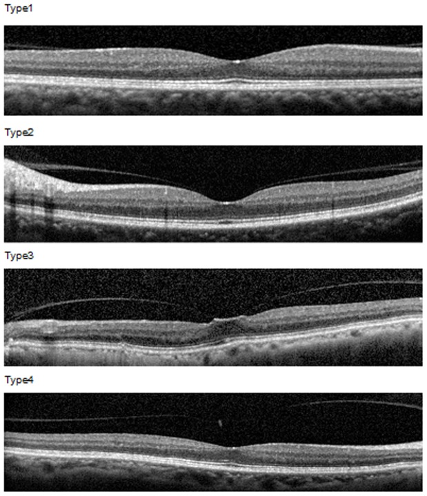
Figure 1. Classification of an incomplete posterior vitreous detachment (PVD).
Type 1: shallow PVD which did not reach the foveal region of the macula; type 2: the PVD reached the fovea but did not extend to the foveola as the center of the fovea; type 3: a shallow PVD with pinpoint foveal vitreous traction on the foveola; type 4: the posterior vitreous was completely detached from the macula including the foveola, but was attached to the optic nerve head.

