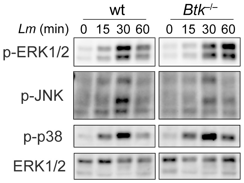
Figure 3. Immunoblot analysis of MAP kinase signaling.

Immunoblot analysis showing the activation of Erk1/2, p38 and JNK1/2 in Wt and Btk −/− BMMs. The cells were infected with Lm (LO28, MOI 10) for the indicated time period. Total ERK1/2 protein levels served as a loading control. Data are representative of three independent experiments. Sustained activation of Erk1/2 has not been observed in the other two batches.
