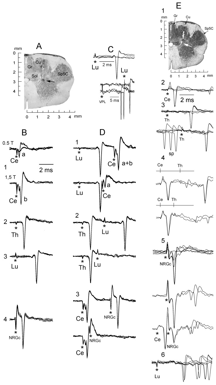
Figure 2. Antidromic identification of spinally-projecting SRD cells collateralizing to or through the NRGc.
A, coronal section showing the approximate site (black arrow) where the silent neuron whose responses are illustrated in B–E was recorded. B, superimposed traces showing the fixed antidromic latency of a cell sending an axon up to the lumbar cord and collateralizing to or through the NRGc. Cervical cord stimulation from subthreshold for cell “b” (B1, lower) revealed the presence of a smaller and shorter-latency spike “a” (B1, upper) elicited solely by this stimulating site. C, all-none response to Lu5 stimulation (upper) and collision between IFP-evoked responses and Lu5-antidromic spikes (lower). D, electrical shocks applied along the cord produced antidromic spikes (upper superimpositions in each panel) that collided at the adequate interstimulus interval (lower superimpositions). Note in D1 (lower) that collision of spike “b” uncovered spike “a”. Stimulus artifacts marked by asterisks. E, a different spontaneously active neuron sending an axon up to the thoracic cord recorded at a site signaled by a white arrow (E1). Successive panels with superimposed records show all-none responses to increasing Ce2 stimulation (E2); all-none response to increasing Th5 stimulation (E3, upper) and collision with spontaneous spikes (E3, lower); Ce2-Th5 collision (E4, lower); all-none response to increasing NRGc stimulation (E5, upper) and Ce2-NRGc collision (E5. Lower); and orthodromic responses to Lu5 stimulation (E6). Cu, nucleus cuneatus; Gr, nucleus gracilis; LRt, nucleus reticularis lateralis; IO, nuclei olivaris inferioris; Py, tractus pyramidalis; Sp5C, nucleus trigeminalis spinalis pars caudalis.

