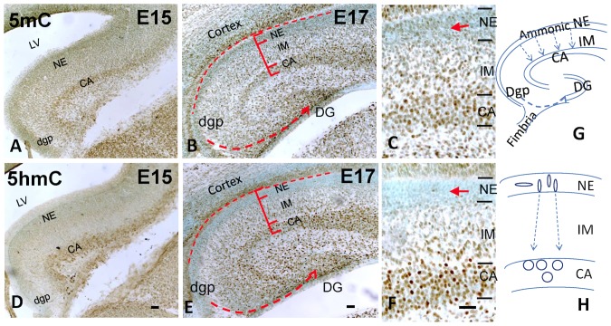
Figure 1. DNA methylation program of developing hippocampus during its early formation from E15 to E17.
Cartoon (G,H) shows the differentiation processes where neuroepithelial (NE) cells migrate into intermediate zone (IM) and arrive in Cornus Ammonis (CA) to become pyramidal neurons; while dentate gyrus neuroepithelium (dgp) migrate through a long journey from lateral hippocampal primodium towards dentate gyrus (DG) and become granule cells. The DNA methylation formed an integral program in these differentiating cells. First progenitor cells all acquire DNA methylation to begin their differentiation. The immunostaining (brown DAB coloration) shows that the 5mC appears ahead of 5hmC as shown in E15 (A,D) and E17 NE (B,E) layer (enlargement, C,F, arrow). There is temporal increment of both methylation marks in CA (pyramidal layer) and developing dentate gyrus (DG) from E15 (A, D) to E17 (B,F). There is also a spatial increment of both 5mC-im and 5hmC-im from NE to IM and to CA at E17 (B,E; higher magnification in C,F, respectively). Moreover, the 5mC-im and 5hmC-im increase as the migration of granule cells from dentate neuroepithelium to the dentate primordial (B,E, dotted line). LV: lateral ventricle. Scale bar: A,B,D,E = 100 µm; C,F = 100 µm.

