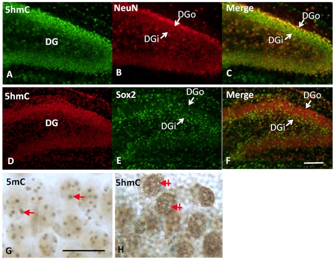
Figure 2. The close association of 5hmC mark with Outside-In pattern of neuronal differentiation in dentate gyrus.
The immunofluorescent double staining reveals that the 5hmC (green in A and C) appeared in maturing or matured neuron as indicated by distribution and co-localization with NeuN (red, B and C), but not in neural progenitor cells marked with Sox2 (green in E and F vs 5hmC red in D and F) in P7 dentate gyrus. When observed through time course, the initiation of neural differentiation is synchronized with the appearance of 5hmC and is demonstrated by co-localization of 5hmC-im with NeuN+ cell in DGo (more mature than DGi), while not with Sox2+ cells in DGi. There is also chromatic translocation of the DNA methylation marks during differentiation, in which the 5mC and 5hmC separate in the nucleus of dentate granule cells (bright field). The 5mC is packed in large granule (G, arrow) and colocalized with DAPI dense area (not shown) in chromatin, while 5hmC is distributed in DAPI-light area complementary to the 5mC+ area (H, cross-arrow indicates 5mC area). DGo: dentate gyrus outer layer; DGi: dentate gyrus inner layer. Scale bar: A–F = 100 µm; G, H = 50 µm.

