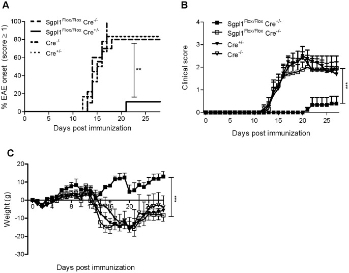
Figure 7. Protection of inducible Sgpl1-deficient mice in EAE.
Tamoxifen-induced Sgpl1Flox/Flox Cre+/−, Sgpl1Flox/Flox Cre−/−, Cre+/−, and Cre−/− mice (n = 6–10/group) were immunized with MOG emulsified in Complete Freund’s Adjuvans. Data from one representative experiment out of three independent studies are shown. A, Incidence of mice with a clinical EAE score ≥1; B, clinical score; C, body weight. For histological analysis thoracic sections of spinal cord tissue from Sgpl1Flox/Flox Cre+/− and Sgpl1Flox/Flox Cre−/− mice undergoing EAE (day 24) were stained (D) with H&E to visualize CNS-invading cells (scale bar is 500 µm, arrows highlight areas of inflammation); E, for CD3+ T cells (scale bar is 500 µm, rectangles indicate area of magnification, where scale bar represents 100 µm); and F, with solochrome to assess the integrity of the myelin sheath (scale bar is 500 µm, arrows highlight areas of beginning demyeliniation).

