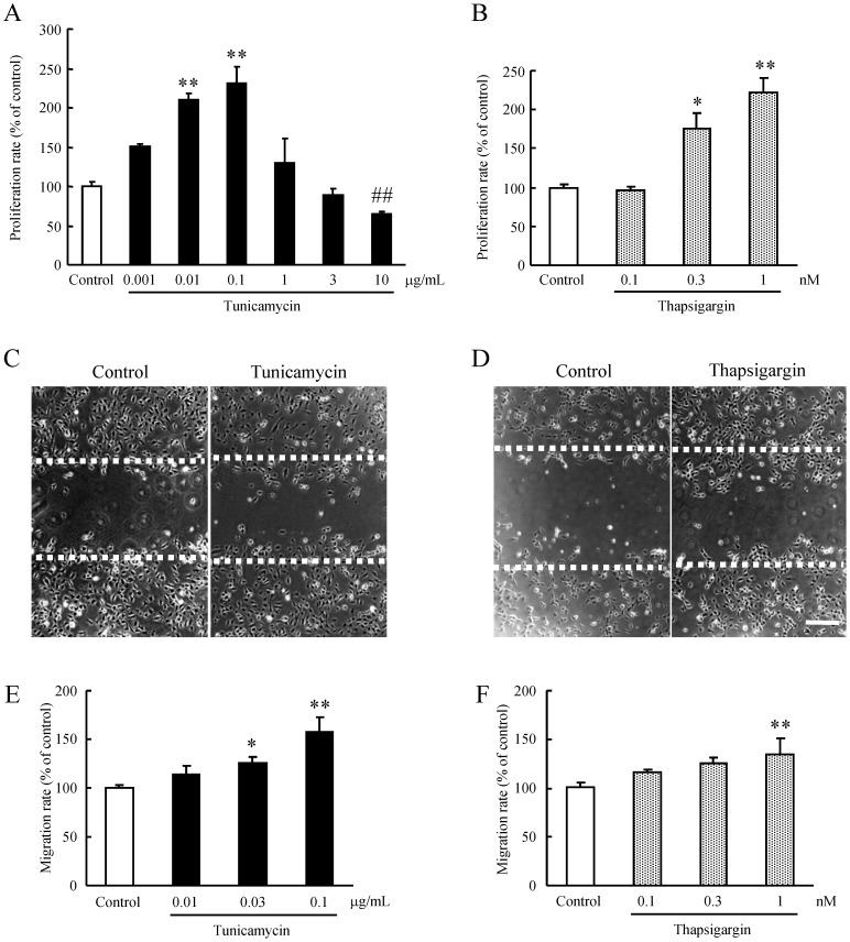
Figure 1. ER stress-induced proliferation and migration in HRMEC.
HRMEC were cultured in a 96-well plate (at a density of 2×10³ cells/well), and were then supplemented with the indicated concentrations of (A) tunicamycin or (B) thapsigargin for 2 h, and measurements were made by WST-8 assay. Data are shown as mean ± S.E.M. (n = 6 or 12). *, p<0.05; **, p<0.01 vs. Control (Dunnett's multiple-comparison test). ##, p<0.01 vs. Control (Student's t-test). Migration of HRMEC was assessed using a wound-healing assay. Briefly, 90% confluent monolayers of HRMEC were scratch-wounded, and then incubated for 24 h. Images of the wounded monolayer of HRMEC were taken at 24 h after treatment for 2 h with (C) tunicamycin or (D) thapsigargin. Migration was estimated by measurement of cell numbers within the wounded region. The indicated concentrations of (E) tunicamycin and (F) thapsigargin increased migration compared to the control. Scale bars indicate 500 µm. Data are shown as mean ± S.E.M. (n = 3 to 7). *, p<0.05; **, p<0.01 vs. Control (Dunnett's multiple-comparison test).

