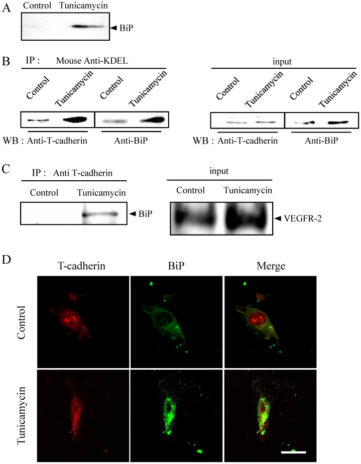
Figure 4. ER stress-induced formation of BiP/T-cadherin complexes on the surface of HRMEC.
HRMEC were treated with 10 ng/ml of tunicamycin for 2 h. (A) BiP on the cell surface was precipitated after biotinylation to detect cell surface protein in untreated (control) or tunicamycin treated cells. (B) BiP and T-cadherin were immunoprecipitated with the anti-KDEL antibody to confirm the association of BiP with T-cadherin. Whole lysate without immunoprecipitation with the anti-BiP antibody was similarly evaluated. (C) After the membrane extraction, BiP and T-cadherin were immunoprecipitated with the anti-T-cadherin antibody and detected by using Western blotting with the anti-BiP antibody. Whole lysate without immunoprecipitation with the anti-VEGF receptor-2 (VEGFR-2) antibody was similarly evaluated to confirm the success of the membrane extraction. (D) Immunostaining showed the presence of BiP and T-cadherin on the surface of HRMEC. Scale bar indicates 30 µm.

