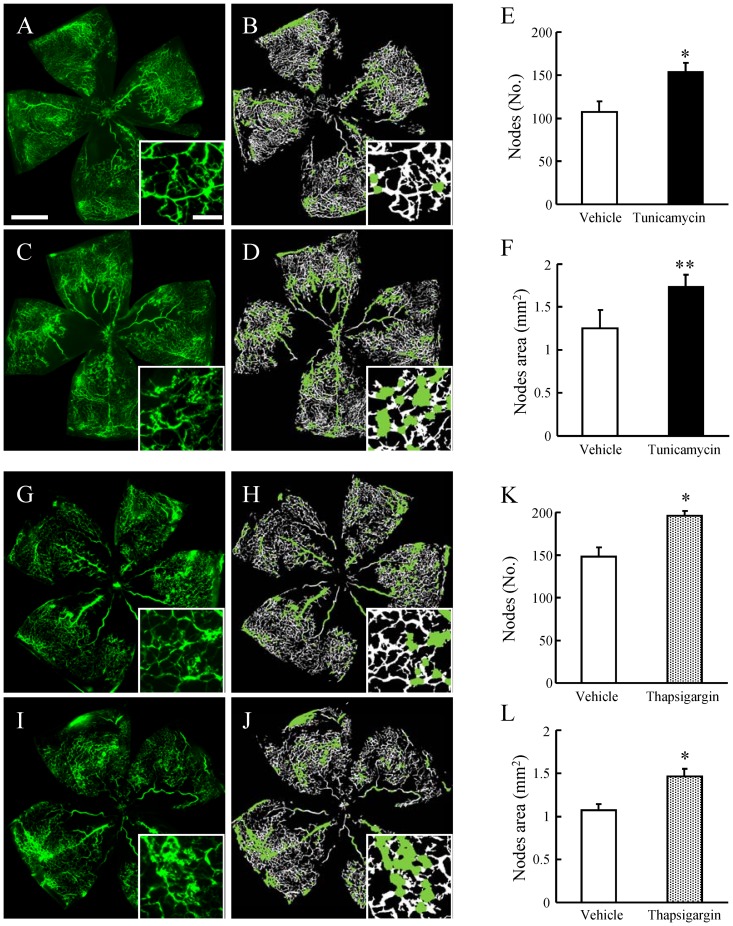
Figure 5. Tunicamycin and thapsigargin accelerated retinal neovascularization in a murine oxygen-induced retinopathy (OIR) model.
The original images (A, C and G, I), together with the analyzed images (B, D and H, J) obtained using the Angiogenesis Tube Formation module in Metamorph, are shown. Scale bars indicate 500 µm and 100 µm (in box). Green labels in the analyzed images show the node regions. Quantitative analysis was performed on the entire retinal microvasculature in flat-mounted retinas obtained at P17. Tunicamycin at 3 µg/ml and thapsigargin at 1 µM increased (vs. vehicle) both the number of nodes (E, K) and the node areas (F, L), which are indexes of pathological neovascularization as calculated using the Angiogenesis Tube Formation module. Data are shown as mean ± S.E.M. (n = 5 to 9). *, p<0.05; **, p<0.01 vs.Vehicle (Student's t-test).

