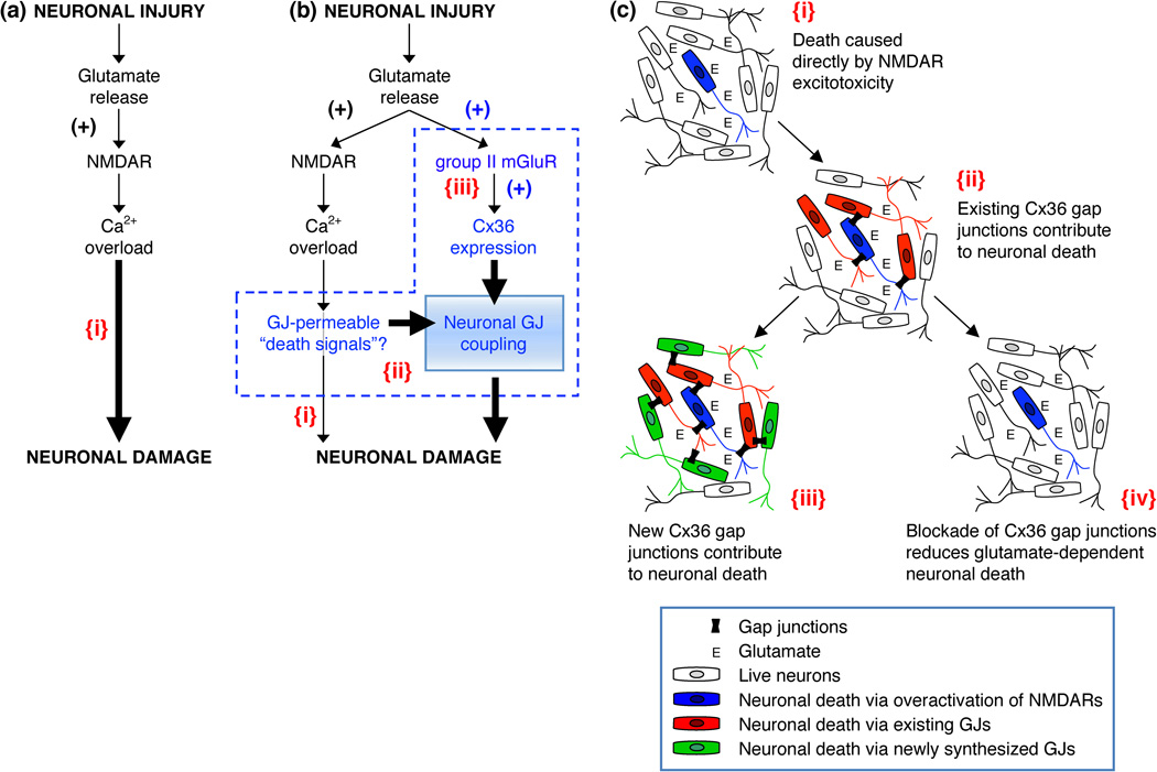
Figure 4. Glutamate-dependent excitotoxicity during neuronal injury.
(a) Traditional model of the mechanisms for glutamate-dependent excitotoxicity [104,105]. (b) Alternative model of the mechanisms of glutamate-dependent excitotoxicity [56]. (c) This figure illustrates key points of the alternative model. In all figures: {i}, Neuronal death caused directly by overactivation of NMDARs; {ii}, Existing neuronal gap junctions (GJs) contribute substantially to neuronal death caused by overactivation of NMDARs [100,109]; {iii}, New neuronal gap junctions are induced by activation of group II mGluRs and also contribute to glutamate-dependent neuronal death [56]; {iv}, Pharmacological [100] or genetic [56,100] blockade of neuronal GJC reduces glutamate-dependent neuronal death. In a,b: the sign (+) indicates the increase in receptor activity or expression of Cx36. In b: a possibility is illustrated that some Ca2+-dependent molecules (or Ca2+ itself) serve as gap junction-permeable death signals (b{ii}) [111]; Ca2+ overload also directly induces neuronal death (b{i}), however, this is not the main driver of neuronal death, rather the key determinant for expansion of cell death is neuronal GJC (b{ii,iii}). Adapted, with permission, from [56] (a and b).

