
Figure 3. Simvastatin reduces brain IFNγ protein expression and brain damage contributed by splenic cells.
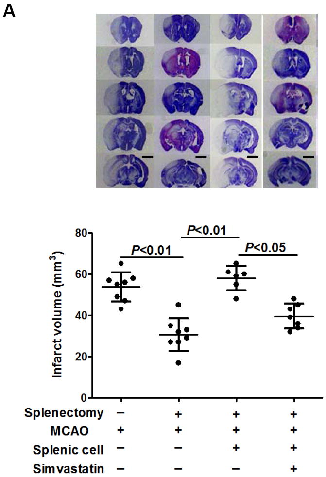
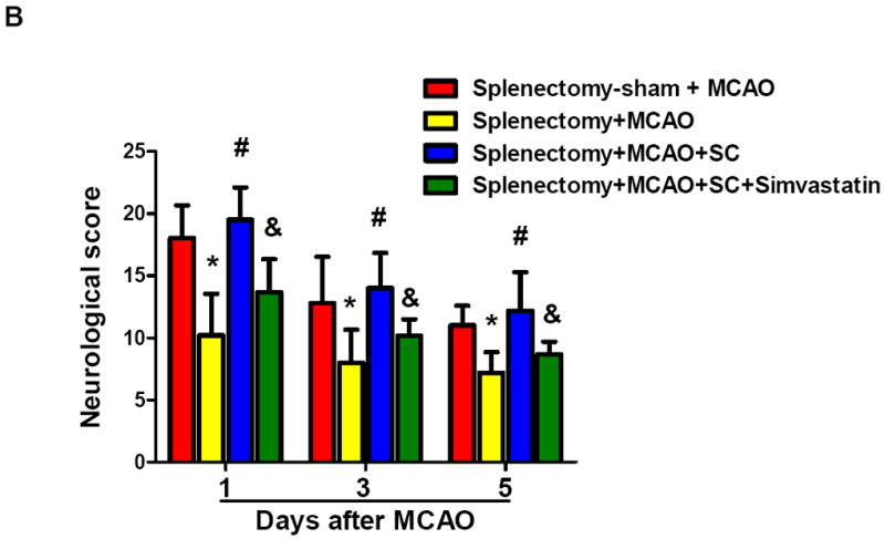
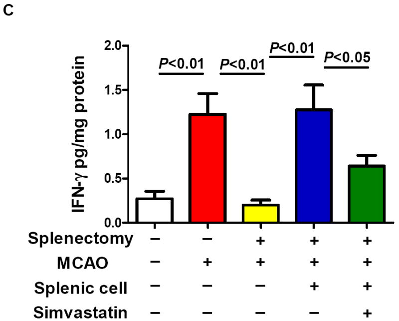
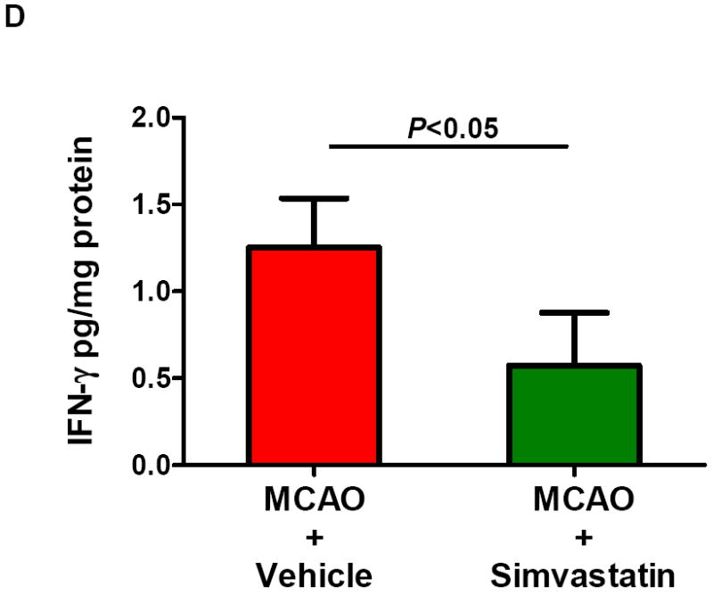
Splenectomy and adoptive transfer of splenocytes were performed as described in the Method. Stroke analysis was performed 5 days after MCAO. A, Representative images of cresyl violet-stained coronal brain sections (upper) and Quantitative analysis of infarct volumes (lower). Scale bar = 2 mm. B, The 28-point neurological scoring at indicated time points (n= 6-8 survived mice/group). *P<0.05 vs. sham-splenectomized MCAO mice; #P<0.05 vs. splenectomized MCAO mice, and &P<0.05 vs. splenectomized MCAO mice with adoptive transfer of splenic cells (SC) at the same time point. C and D, ELISA measurement of IFN-γ protein concentrations in the ipsilateral hemispheres from the indicated groups at 72h after MCAO. n=5 mice/group.