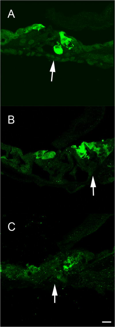
Figure 4.

Delivery of Ad28.gfap.atoh1 results in development of auditory hair cells. Temporal bones were evaluated for the presence of hair cells one month after delivery of atoh1 by immunofluorescent staining for myosin VII. In the apical turn of treated (left) ears, myosin positive staining can be seen in both the inner and outer hair cell regions (A). The arrow indicates a myosin VII positive cell in Corti’s tunnel below a normal appearing inner hair cell. Two myosin VII positive cells can be seen lateral to the normal outer hair cell region. In the midturn of the treated side (B), multiple myosin VII positive cells are seen in the inner hair cell region (arrow) indicating the presence of supernumerary hair cells. Three myosin VII positive cell that do not extend to the basement membrane can be seen in the outer hair cell region of the midturn organ of Corti. In the untreated ear only minimal myosin VII positive debris can be seen in the apical turn (C), with no indication of normal hair cells in any of the turns.
