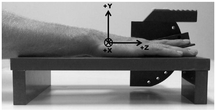
Figure 1.

Schematic of device used to position index finger during MRI scans. Included is the coordinate system of the hand/forearm using the hook of hamate as the origin. The Z-axis was aligned with the longitudinal axis of the forearm with the proximal to distal direction being positive. The Y-axis was orientated in the anterior to posterior (positive) direction and the X-axis was oriented in the lateral to medial (positive) direction.
