Fig. 12.

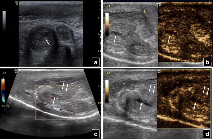
Transverse B-mode scan of the penis of a 36-year-old man (a) shows enlargement of the right corpus cavernosum penis, as well as an indistinct hypoechoic area (arrow) in its centre. This area shows no contrast enhancement on CEUS (arrow in b) and is consistent with an injury. Sagittal colour Doppler scan (c) also shows this traumatic area (arrow), while loss of surrounding fibrous tissue intactness (double arrows) is suggested. The findings of corpus cavernosum injury (arrow) and surrounding tissue rupture (double arrows) are confirmed in the sagittal CEUS view (d)
