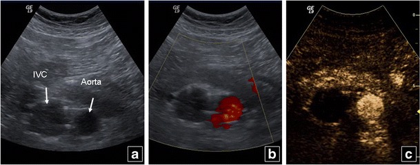
Fig. 13.

A 74-year-old male patient with bilateral leg deep venous thrombosis history and IVC filter. B mode US (a) shows a dilated IVC, while no blood flow is noted on colour Doppler (b). IVC thrombosis is confirmed on CEUS (c) with absence of contrast in the IVC, contrary to the normally enhancing abdominal aorta
