Fig. 9.

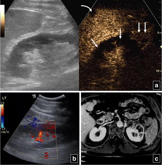
Inhomogeneous echogenicity is noted on B mode US (left part of a) of the left kidney in a 56-year-old man. Colour Doppler US (b) is suboptimal for the detection of blood flow in the renal cortex. CEUS (right part of a) shows absence of flow in parts of the cortex due to partial infarction (arrows), while other parts of the cortex (double arrows) and medulla enhance normally. A small splenic infarct (curved arrow in a) is also seen on CEUS. Left kidney partial cortical infarction is confirmed on MR (c). The splenic infarction is not seen at this level
