
Fig. 5.
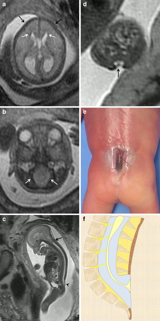
Myeloschisis. Fetus at 23 weeks’ gestation. a, b Axial T2-weighted HASTE image of the fetal cranium shows the deformity of the frontal bones (black arrows in a) and the characteristic beaklike shape of the ventricular horns (arrows in a). The posterior fossa deformity can also be seen: the cisterna magnum is absent and the foramen magnum is enlarged due to the descent of the structures of the posterior fossa toward the spinal canal (arrows in b). c Sagittal T2-weighted HASTE image shows a cutaneous defect or thinning in the lumbosacral region (arrowhead) with no mass protruding through the defect as well as an associated Chiari malformation (black arrow). d Axial image at the level of the cutaneous defect shows the abnormally low neural placode within the spinal canal without expansion of the subarachnoid spaces. These findings are characteristic of myeloschisis. e Pathological specimen. f Sagittal diagrams of the anomaly