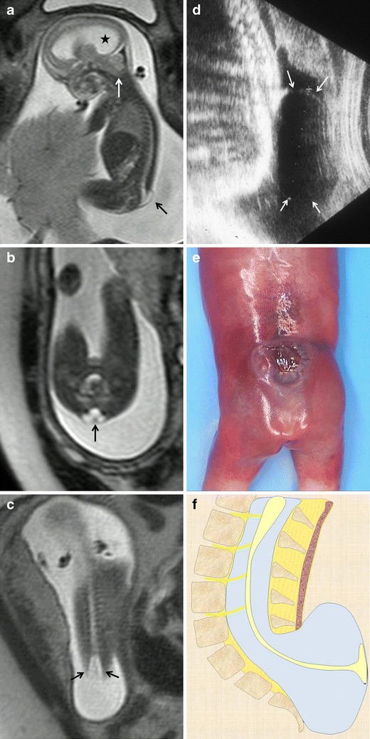
Fig. 6.

Lumbosacral myelomeningocele. Fetus at 22 weeks’ gestation. a Sagittal T2-weighted HASTE image shows a small cystic mass protruding at the thoracolumbar level (black arrow). Associated hydrocephalus (asterisk) and a Chiari malformation (white arrow) are also seen. b Axial T2-weighted HASTE image at the lumbosacral level shows the neural placode protruding outside the surface of the skin due to expansion of the adjacent subarachnoid space (black arrow). c Coronal image at the spinal level shows increased interpedicular distance at the level of the neural defect. d Ultrasound image of a different patient shows a cystic mass protruding at lumbar level (white arrows). e Pathological specimen. f Sagittal diagrams of the anomaly
