Fig. 7.

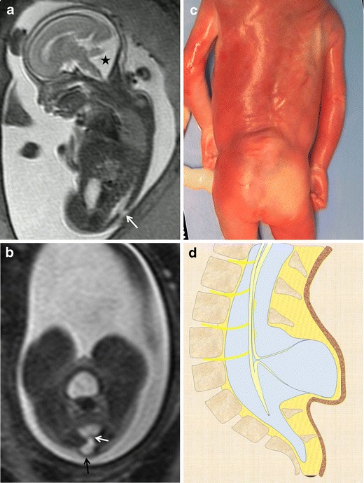
Myelocystocele. Fetus at 23 weeks’ gestation. a Sagittal T2-weighted HASTE image shows an interruption in the posterior vertebral arches in the lumbar region (arrow). A posterior fossa anomaly with an elevated vermis and dilation of the IV ventricle is also seen (asterisk). b Axial image at the lumbar level showing the cystic mass protruding through the spinal defect (black arrow). Two nerve roots (white arrow) can be seen arising from the non-neurulated neural placode inside the spinal defect. c Pathological specimen. d Sagittal diagram of the anomaly
