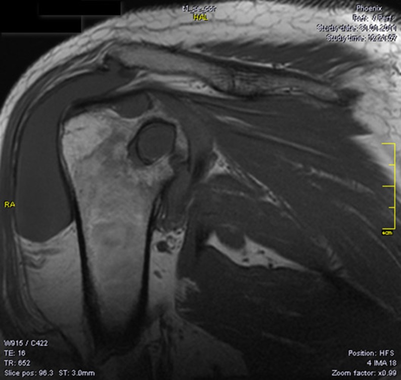
Fig. 3.
Magnetic resonance imaging (MRI) of the shoulder demonstrates synovial hypertrophy, diffuse oedema of the humeral head and of the glenoid, small marginal erosions and large subchondral cysts on both articular surfaces, larger on the superior part of the humeral head (22 mm), effusion in the subacromial bursa

