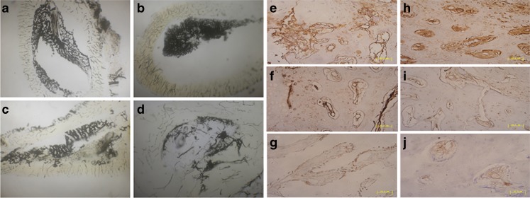
Fig. 4.
Morphological and histological observation. Axial view (4× magnification) of Chinese-ink microangiographs at 12 weeks post operation showed that the cross-sectional morphology of the medullary cavity and intraosseous vasculatures, including intramedullary blood vessels in group A (b) was restored close to normal (a). However, the medullary vessels and medullary cavity were partly restored in group B (c) and barely rebuilt in group C (d). Early vascularization was enhanced by EPC prevascularization with formation of neovasculature expressed higher VEGF (e) and FVIII (h) immunopositivity at two weeks. In group B, the number of neovasculature was less than group A, and the expression of VEGF (f) and FVIII (i) was moderate positive. Few blood vessels with weakly positive expression of VEGF (g) and FVIII (j) could be observed in group C; bar length = 50 μm

