
Figure 8.
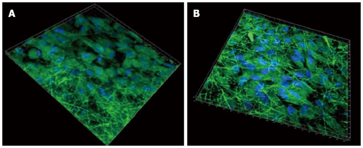
3D image using Imaris software of cardiomyocytes-mesenchymal stem cells co-culture group stained with cardiac specific marker protein troponin at 60 × magnification on (A) collagen fibers (B) poly (glycerol sebacate)/collagen core/shell fibers. Nucleus stained with 4,6-diamidino-2-phenylindole hydrochloride.