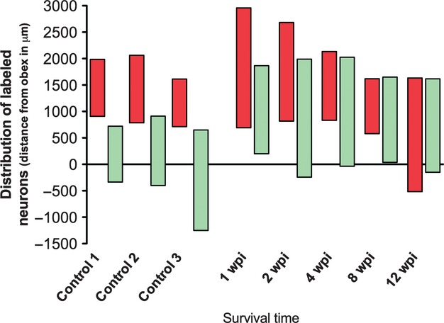
Fig. 3.

Histogram showing the rostrocaudal position of labeled motoneurons present in control animals and in experimental animals allowed to survive for different times following laryngeal nerve crush injury. Labeled motoneurons after the injection of tracer in the PCA muscle appear in red, and in green those of the TA muscle. The 0 represents the obex, the positive values represent the rostral distance from the obex, and the negative values the caudal distance from the obex (wpi, weeks post-injury).
