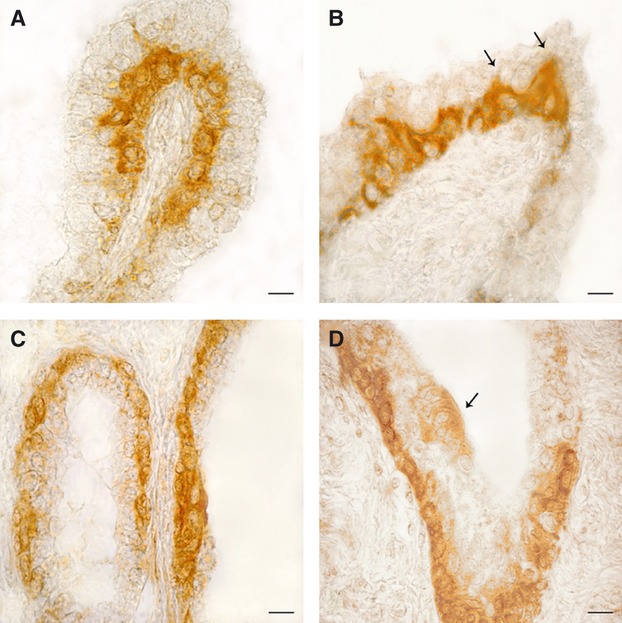
Fig. 2.

OXA- (A and B) and OX1R- (C and D) immunoreactivity in the human hyperplastic prostate. (A) The basal membrane of an intrafollicular, papillar-like structure is lined by a continuous row of intensely stained cells. (B) A peculiarity often shown by positive basal cells is the presence of a slender cytoplasmic extension (arrows) directed towards the follicular lumen and intermingled between the negative apical cells. (C) Almost all basal cells of the follicular epithelium contain OX1R-immunoreactive material, which completely fills their cytoplasm. (D) Invagination of a prostatic follicle completely lined by positive basal cells. The arrow points to a small cluster of low intensely stained apical cells close in contact with the follicular fluid. Avidin–biotin immunohistochemical method. Scale bars: 20 μm.
