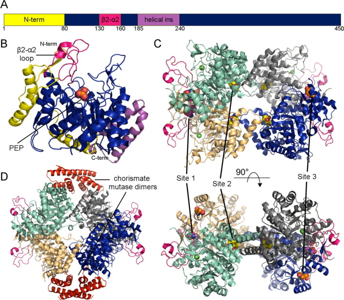
Figure 4.

Overview of type II DAHPS structure and allostery. (A) Schematic representation of type II domain construction highlighting the three most prominent insertions (B) The M. tuberculosis DAHPS (PDB code 2B7O) monomer highlighting the characteristic type II inserts. (C) The type II DAHPS (PDB code 2YPQ) tetramer complexed with tryptophan (site 1, yellow) + phenylalanine (site 2, purple and site 3). The active site manganese ion is depicted as a green sphere. (D) Structure of the M. tuberculosis DAHPS + chorismate mutase (red) complex (PDB code 2W19). An interactive view is available in the electronic version of the article.
