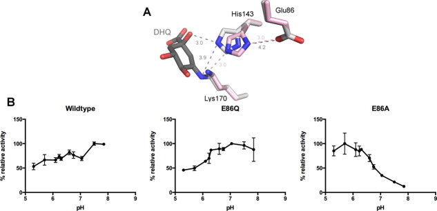
Figure 1.

(A) Superposition of seDHQD unliganded (pink, PDB code: 3L2I) and bound DHQ reaction intermediate (gray, PDB code: 3M7W) structures, displaying the putative catalytic triad: Glu86, His143, and Lys170. Distances are shown in angstroms and colored by structure. (B) Maximal DHQD activity by pH of wild-type, E86Q, and E86A variants. Activity is normalized to the most active pH for each variant. Standard deviations are indicated as error bars.
