
Figure 2.
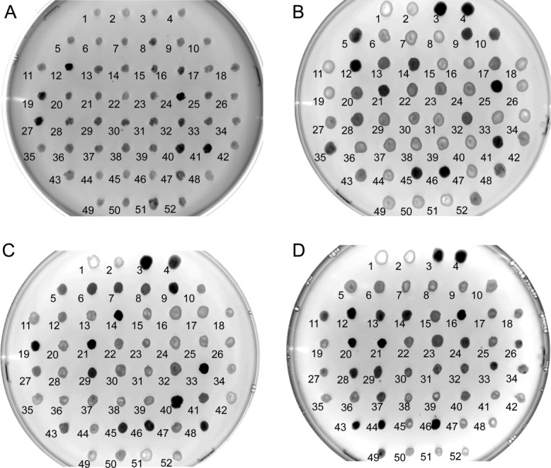
Induction plate expression screening. (A) AQP4, (B) human PEMT, (C) mouse PEMT, and (D) yeast PEMT (OPI3). Clones were spotted onto BMMY plates and incubated for 24 h at 30°C. P. pastoris GS115 was spotted onto position 1 and 2 as a negative control. Well expressing clones of AQP4 (clones 25, 42) were included as positive controls in positions 3 and 4 on plates B–D. Plates were imaged under blue light using an ImageQuant LAS 4000 with a 1/8th of a second exposure.