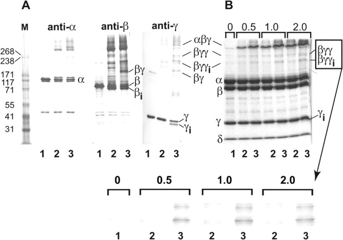
Figure 8.

Mg2+-dependent crosslinking of PhK with dibromobimane at pH 6.8. (A) Native PhK (Lane 1—control) was crosslinked with dibromobimane for 1.0 min following incubation for 2.0 min in the absence (Lane 2) and presence (Lane 3) of Mg2+. The crosslinking was quenched by addition of SDS buffer. Following electrophoresis, all conjugates were identified by their apparent masses and cross reactivities against subunit-specific mAbs using previously described methods.57 No conjugates containing the intrinsic calmodulin (δ) subunit were detected in Western blots with the anti-calmodulin mAb (data not shown). Subunits and all conjugates formed by crosslinking are indicated both to the left and right of the blots and gel, with intramolecular crosslinking of species indicated by subscript i. (B) Crosslinking and electrophoresis of PhK were carried out as above, but then the proteins were stained directly in gel with Coomassie blue. Prior to crosslinking for 1 min, PhK was incubated (± Mg2+) for times varying between 0.5 and 2.0 min (indicated numerically above gel). Species that underwent Mg2+-dependent crosslinking are indicated to the right of the gel. The Mg2+-dependent formation of βγγ and βγγi is shown expanded below the gel. Masses for standard markers (Column M) are indicated in kDa to the left of the blots and gel.
