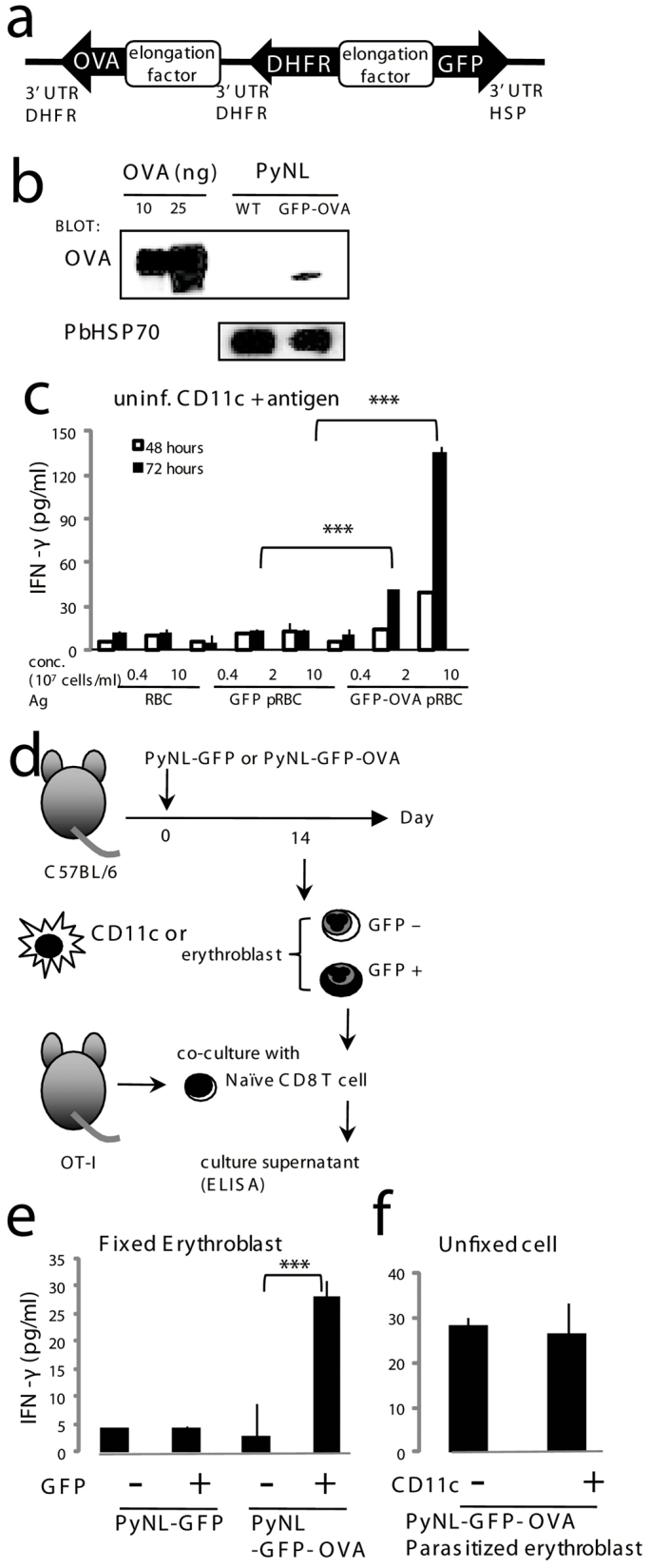
Figure 7. Parasitized erythroblasts activate CD8+ T cells.

(a) Construction of the Plasmodium artificial chromosome (PAC) containing GFP and OVA. PyNL parasites were transfected and the recombinants selected based on resistance to pyrimethamine conferred by exogenous expression of DHFR. (b) Expression of OVA in the recombinant parasite was detected by western blot analysis. Protein extracts from parasitized RBCs purified from mice infected with the recombinant and parental (WT) parasite and OVA protein were separated on SDS-PAGE and transferred to nitrocellulose membranes followed by blotting with an anti-OVA antibody. An antibody against P. berghei HSP70 was used as an internal control. (c) Immunogenicity of OVA-expressing recombinant parasites. Purified CD11c+ cells (2 × 10⁴) from uninfected mice were co-cultured with 105 OT-I CD8+ T cells in the presence of antigens from the indicated cells. (d) Protocol for antigen-specific recognition of parasitized erythroblasts. GFP+ parasitized or GFP− uninfected erythroblasts isolated from spleen samples of C57BL/6 mice infected with PyNL-GFP or PyNL-GFP-OVA as shown in Fig. 4b. (e) Purified CD11c+ cells (2 × 10⁴) from C57BL/6 mice infected with the indicated parasite were pulsed with or without OVA peptide and fixed, then were co-cultured with 105 OT-I CD8+ T cells. (f) Purified PyNL-GFP-OVA parasitized erythroblasts (2 × 10⁴ cells) were mixed with or without CD11c+ cells (2 × 10⁴) from PyNL-GFP-OVA infected mouse and co-cultured with purified CD8+ CD11c− T cells (105 cells) from Rag2−/− OT-I mice. (c, e, f) Activation of OT-I CD8+ T cells was assessed by production of IFN-γ. Culture supernatants were collected after 72 hours otherwise mentioned, and the concentration of IFN-γ was measured. Mean ± S.D. from triplicate cultures from 4 mice is shown. Representative data from two to six independent experiments are shown. Statistical significance is indicated as *P < 0.05 and ***P < 0.001.
