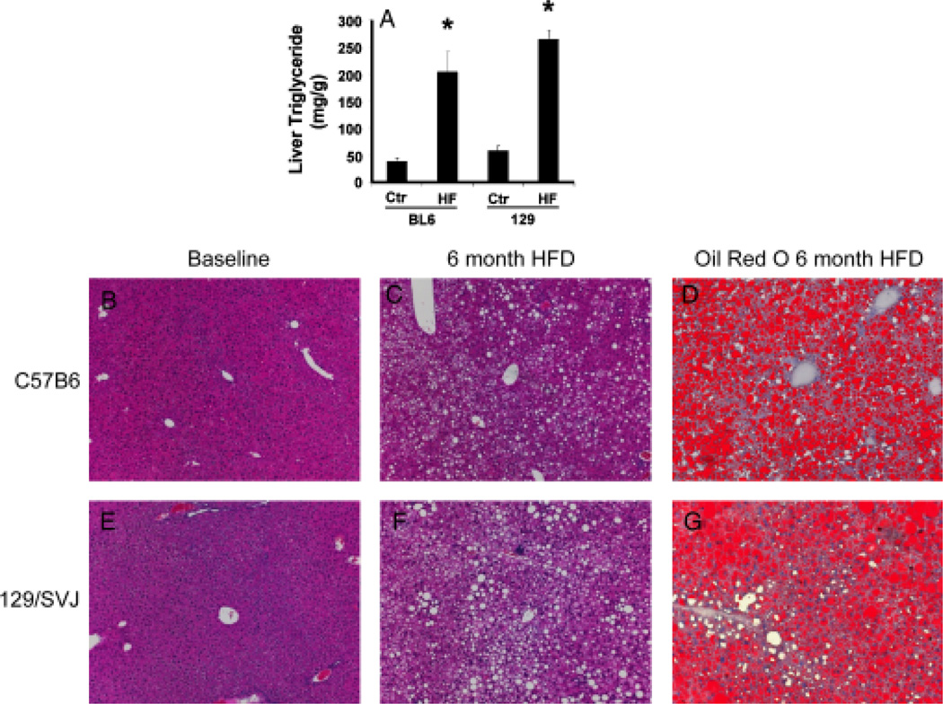
Fig. 1.
C57BL6 mice and 129/SVJ mice developed similar degrees of hepatic steatosis after 6 months of high-fat (HF) diets. Hepatic triglyceride content in C57BL6 mice (BL6) and 129/SVJ mice (129) after ingesting regular chow (control, Ctr) (n = 11/group) or HF diet (n = 26/group) for 6 months (A). Histology in representative mice (B–G). Haematoxylin and eosin-stained liver sections in BL6 mice (B, C) and 129/SVJ mice (E, F) at baseline (B, E) and after 6 months of HF diets (C, F). Oil red O staining demonstrates neutral lipids in livers of BL6 mice (D) and 129/SVJ mice (G) after 6 months of HF diets. *P<0.05 vs Ctr.

