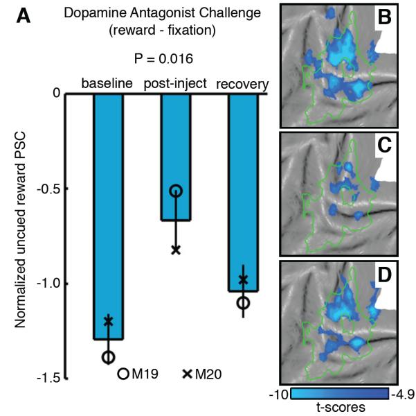
Figure 8. Uncued reward activity in visual cortex is susceptible to dopamine D1-receptor antagonist (SCH-23390) challenge (experiment 6).
(A) Mean normalized PSC (see Supplemental Experimental Procedures) during uncued rewards (uncued reward – fixation, group-level analysis, 30 runs/phase, M19 & M20 - 15 runs/phase) within the cue-representation (see Table S1) measured during baseline, post-injection and recovery phases. Error bars denote SEM across runs. Symbols denote the mean normalized PSC of single-subject analyses [M19 (circle); M20 (cross)]. Kruskal-Wallis non-parametric ANOVA was performed comparing PSC across phases. Uncued reward fMRI activity (uncued reward – fixation, P < 0.05 FWE corrected group-level analysis, 30 runs/phase, M19 & M20 - 15 runs/phase) projected onto a flattened cortical representation of left occipital cortex during the (B) baseline, (C) post-injection, and (D) recovery phases. Green outline represents the cue-representation. See also Table S7 and Figure S8.

