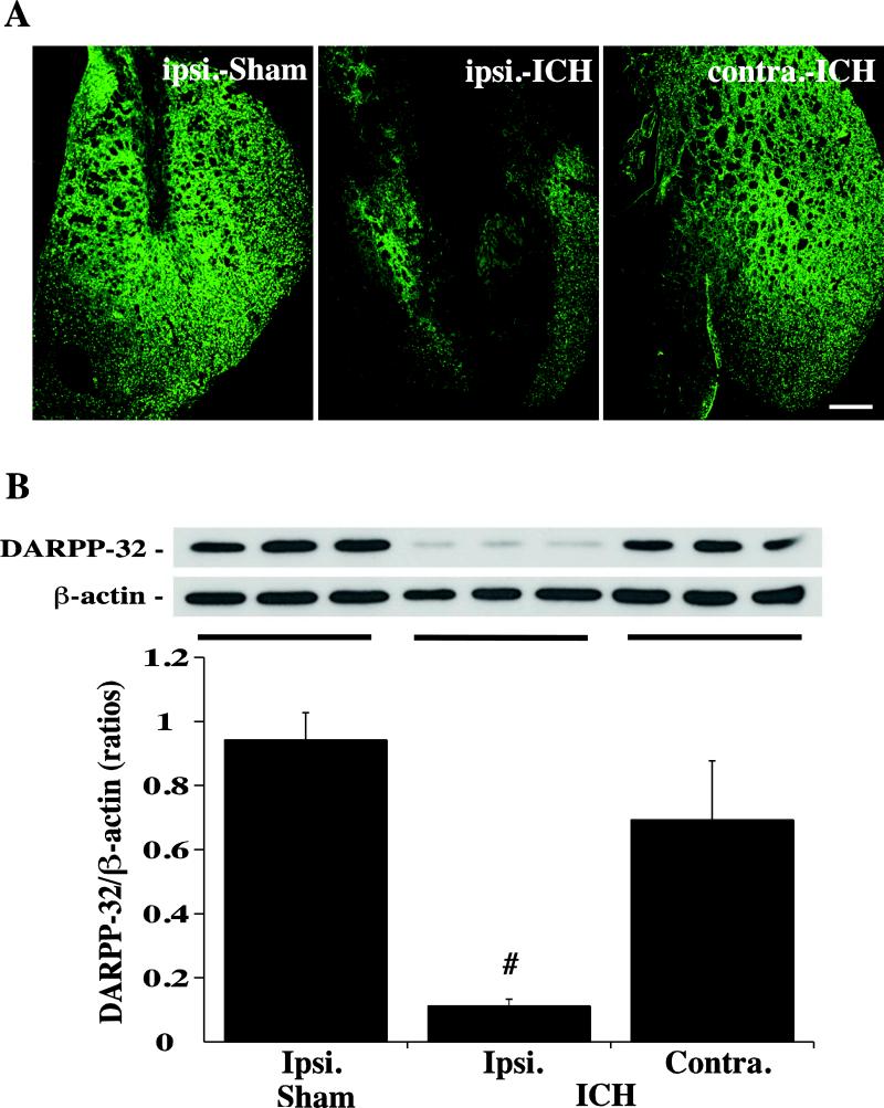
Figure 3.
Immunoreactivity (A) and protein levels (B, Western blot) of DARPP-32 in the ipsi- and contralateral basal ganglia at day-3 after a needle (Sham) or 100μl blood (ICH) injected into the right caudate. β-actin was examined as a protein loading control for Western blots and DARPP-32 levels expressed a ratio to β-actin levels. Values are means ± SD, n=3, # p<0.01 vs. in Sham and in contralateral. Scale bar = 500μm.

