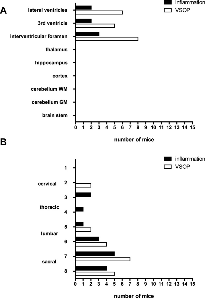
Figure 1. Histological detection of inflammation and VSOP prior to EAE clinical onset (4–6 days post transfer).
The number of mice showing inflammation, defined as pathological accumulations of immune cells as revealed by H&E staining, and VSOP as shown by Prussian Blue staining, is indicated for various locations in the brain (A). The spinal cord (B) was cut into eight transverse segments spanning from the cervical to sacral zones. n=15.

