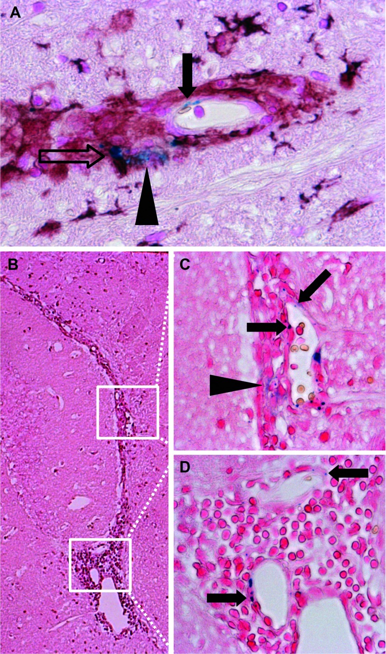
Figure 7. VSOP present in multiple forms in CNS lesions at peak disease.
VSOP could be seen as discrete puncta that appear to be present in elongated endothelial structures (solid arrow in A). VSOP was also seen in the same lesion co-localized with an iba-1-positive cell (open arrow in A), and as a diffuse accumulation (arrowhead in A). Multiple forms of VSOP in inflamed choroid plexus in the interventricular foramen (B). VSOP as discrete puncta structures (solid arrows in C and D), and as a diffuse accumulation (arrowhead in C). Original magnification: (A, C, D) ×200; (B) ×100.

