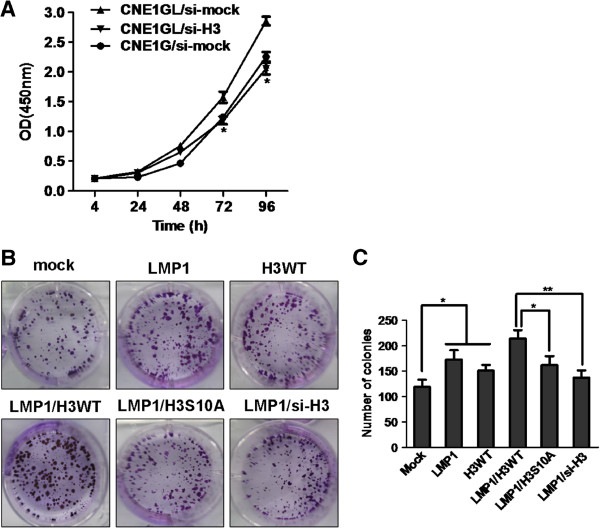
Figure 3.
Phosphorylation of histone H3 at Ser10 was involved in LMP1-induced CNE1 cell transformation. (A) CNE1G and CNE1GL cells were transfected with si-mock or si-H3 and then cell proliferation was estimated at 24-hour intervals up to 96 hours using CCK-8 assay. Data were presented using mean±SD. Asterisks indicate a significant difference compared with CNE1GL/si-mock control cells (*, p < 0.01). (B) and (C) CNE1 cells transfected with various combinations of expression vectors as indicated were subjected to a focus-forming assay. The cells were cultured for 2 weeks and foci were stained with 0.5% crystal violet. The average foci number was calculated and presented using mean±SD. Asterisks indicate a significant difference in foci formation between indicated groups. (*, p < 0.05; **, p < 0.005).

