Fig. 1.
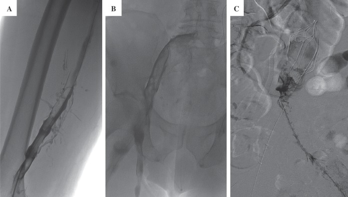
Extensive thrombus from the inferior vena cava (IVC) to the popliteal vein.
A: Femoropopliteal venogram at day #0 reveals extensive thrombus.
B: Iliofemoral venogram at day #0.
C: Venogram at day #1 demonstrates extensive amounts of caval thrombus.
Note the IVC filter. An AngioJet® mechanical thrombectomy catheter (Possis Medical, Minneapolis, MN) was used to help clear the clot burden in the IVC.

