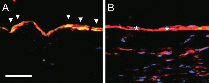
Fig. 4.

Vein graft adaptation in rats. Photomicrographs of jugular vein (A) and vein graft explanted at 21 days (B) in rats, processed for immunofluorescence to detect Eph-B4 (green), the smooth muscle cell marker α-actin (red), and the nuclear marker DAPI (blue). Scale bar, 20 microns. Arrowheads show colocalization of Eph-B4 with α-actin in the vein, consistent with the presence of Eph-B4 in venous smooth muscle cells; stars show loss of colocalization and Eph-B4 in the vein graft, with persistent detection of the smooth muscle cells.
