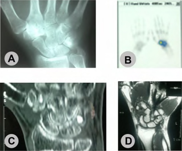
Fig. 3.
Osteoid osteoma of trapezoid bone of 31 years old men with wrist pain since 18 month ago. A: Anteroposterior radiography shows sclerosis at trapezoid bone. B: Bone scan shows increased local uptake. C: CT scan shows nidus at trapezoid (incidental bone iland at triquetrum). D: The MRI shows diffuse edema at wrist.

